Pathologists partner with community physicians to diagnose difficult cancer cases
Pathologists are physicians who perform laboratory studies to diagnose disease, but their role is especially important in oncology. Cancer, more than any other disease, is difficult to diagnose precisely. Subtle differences in the size or shape of a tumor, its genetic makeup or the types of molecules on the surface of cancer cells can make a huge difference in identifying the right treatment.
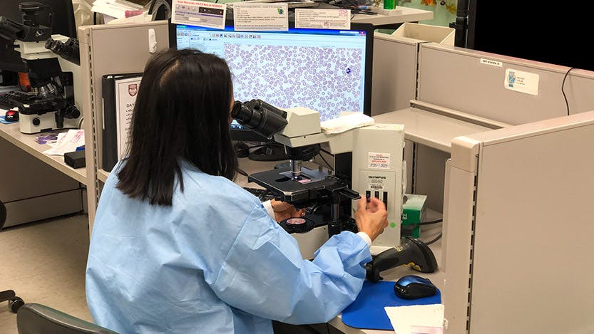
Thirty years ago, pathologists spent much of their time looking through a microscope, scrutinizing tissue or blood samples on a glass slide for the misshapen cells and tiny discrepancies that might give away a malignancy or define a certain sub-type of tumor. Today, the microscope is still an indispensable tool, but pathologists also have access to a vast array of modern technologies to diagnose cancers: special stains and dyes to see genetic mutations, various molecular tests including next generation genetic sequencing, advanced cellular imaging and precise blood testing techniques. These new technologies have redefined the field, but the pathologist’s mission remains the same: making a precise diagnosis to find the right treatment for each patient.
The University of Chicago Medicine has a robust pathology department staffed by leading experts equipped with the latest versions of these tools, but not all patients start their medical journey at a large academic medical center. Instead, many of them visit a community hospital or their family physician closer to home. Smaller hospitals have their own pathology services, but they may not be able to perform all the necessary tests or have the expertise to make a definitive diagnosis, so they often refer these cases to larger facilities.
Partnering with community physicians
UChicago Medicine also has a growing pathology consult service to assist with these referrals or to provide second opinions on diagnoses patients have received elsewhere. Daniel Arber, MD, the Chair of the Department of Pathology, says that these services build strong working relationships with community physicians who refer difficult cases for help, but usually continue treating the patients once they have a definitive diagnosis.
“What we do is partner with them, we don't want to take the patients away,” Arber said. “They'll send us the slides and tissues if we need to do more tests and we send the report back to them. It helps them with their practice, and it’s more interesting for us because usually they’re difficult cases.”
What we do is partner with them, we don't want to take the patients away ... It helps them with their practice, and it’s more interesting for us because usually they’re difficult cases.
Peter Pytel, MD, a neuropathologist who specializes in examining nervous system tissues, brain tumors and skeletal muscle, said the additional services they can provide are a matter of resources and scale.
“It’s not that we are better pathologists, but we have greater opportunities with a lab that might offer additional tests not available to these other facilities to back up ideas about possible diagnoses,” he said.
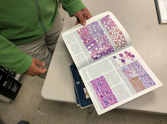
For Pytel, that usually starts by working with tumor samples encased in paraffin wax blocks. These can be sliced into thin sections that can be used for special stains highlighting otherwise unrecognizable aspects of the tissue or can be used to extract DNA and RNA for genetic studies. Some samples like nerve and muscle biopsies are triaged and stored in special ways that may include frozen tissue and a separate set of unique special stains.
“These are specialized services and use different techniques than those used in a general pathology lab,” Pytel said. “It’s often not worth it for them to set up all the triage required for these muscle and nerve biopsies.”
New technology and specialized services
Such specialized services are also particularly important to hematopathologists who focus on diagnosing blood disorders and cancers like lymphoma or leukemia. Sandeep Gurbuxani, MD, PhD, works next to a bustling laboratory filled with high-volume machinery to process blood, urine and tissue samples from the entire hospital.
A portion of that equipment is dedicated to advanced technology for diagnosing cancer. For example, flow cytometry is a technique that looks for specific antigens, or molecules expressed on the surface of cancer cells. Different types of cancers express different antigens, and flow cytometry uses lasers to detect fluorescent tags that are embedded in antibodies that bind to them. This can help identify exactly what kind of cancer is present, which in turn determines the appropriate treatments. Similarly, immunohistochemistry identifies antigens and other proteins in a cell by using enzymes or dyes to stain these molecules so they can be seen under a microscope.
Pathologists also rely increasingly on genetic sequencing to diagnose cancers. Cancer is a genetic disease at its core, caused by mutations or mistakes in the way genes are used which lead to changes in the way cells grow and divide. Modern genetic sequencing tools have vastly lowered the cost and amount of time needed to analyze the DNA in tissue and blood samples and identify the genetic anomalies that mark various sub-types of cancer.
“About 30 years ago, the glass slides would’ve been the one tool consistently available to us for a workup,” Gurbuxani said. “We are now at the point where it’s just not enough to look at microscopic changes. As we have advanced our understanding of the genetics of tumors we have started to rely on these next generation sequencing techniques to fine tune our diagnoses and provide targets for treatment,” he said.
Deep expertise beyond the textbook
Nicole Cipriani, MD, is a surgical pathologist who specializes in both head and neck pathology as well as bone and soft tissue pathology. She says that the role pathologists at UChicago play when reviewing cases on consultation from other hospitals is to provide a fresh set of eyes.
“The best practice is to look at them almost blinded, without knowing what the outside hospital’s diagnosis was because that helps me treat it as if it were my own case,” she said.
Cipriani is also working to launch a “virtual pathology” service at UChicago Medicine that will digitize all pathology slide images so they can be viewed on a computer. This will establish a secure network that allows pathologists and treating physicians to review slides together from any location, and seamlessly transfer images and data back and forth when it isn’t feasible to ship physical samples or tissue blocks to Chicago for review.
Cipriani stressed the complexity of cases that end of being referred to UChicago as a means of building deep expertise in their respective sub-specialties. The more generalized pathology departments at smaller hospitals don’t have trouble diagnosing tumors that fit the textbook definition of a certain type of cancer. Rather, they turn to UChicago for help, as Cipriani said, “when the tumor didn’t read the book.”
Most patients may not realize what pathologists do, but they play a seminal role in their care. Pytel says a certain type of doctor is drawn to pathology, one with more of an interest in basic science who is comfortable working in the background. While they may not get the reward of treating a patient through their medical journey, the work pays off in different ways.
“In pathology, we see our patients through the slides,” he said. “But because it’s such a brief encounter, we can see many more patients. That allows us to see more of the outlier cases, the unusual and rare cases that most doctors only get to learn about in medical school.”